1. Signaling Pathways
  2. Apoptosis
  3. TNF Receptor

TNF Receptor (肿瘤坏死因子)

Tumor Necrosis Factor Receptor; TNFR

Tumor necrosis factor (TNF) is a major mediator of apoptosis as well as inflammation and immunity, and it has been implicated in the pathogenesis of a wide spectrum of human diseases, including sepsis, diabetes, cancer, osteoporosis, multiple sclerosis, rheumatoid arthritis, and inflammatory bowel diseases.

TNF-α is a 17-kDa protein consisting of 157 amino acids that is a homotrimer in solution. In humans, the gene is mapped to chromosome 6. Its bioactivity is mainly regulated by soluble TNF-α–binding receptors. TNF-α is mainly produced by activated macrophages, T lymphocytes, and natural killer cells. Lower expression is known for a variety of other cells, including fibroblasts, smooth muscle cells, and tumor cells. In cells, TNF-α is synthesized as pro-TNF (26 kDa), which is membrane-bound and is released upon cleavage of its pro domain by TNF-converting enzyme (TACE).

Many of the TNF-induced cellular responses are mediated by either one of the two TNF receptors, TNF-R1 and TNF-R2, both of which belong to the TNF receptor super-family. In response to TNF treatment, the transcription factor NF-κB and MAP kinases, including ERK, p38 and JNK, are activated in most types of cells and, in some cases, apoptosis or necrosis could also be induced. However, induction of apoptosis or necrosis is mainly achieved through TNFR1, which is also known as a death receptor. Activation of the NF-κB and MAPKs plays an important role in the induction of many cytokines and immune-regulatory proteins and is pivotal for many inflammatory responses.

Cat. No. Product Name Effect Purity Chemical Structure
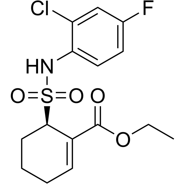
  • HY-11109
    Resatorvid

    瑞沙托维

    Inhibitor 99.95%
    Resatorvid (TAK-242) 是一种选择性的 TLR4 信号传导抑制剂。Resatorvid 抑制 NOTNF-αIL-6 的产生,其 IC50 值分别为 1.8 nM,1.9 nM,1.3 nM。Resatorvid 下调 TLR4 下游信号分子 MyD88 和 TRIF 的表达。Resatorvid 抑制自噬 (autophagy),并在各种炎症性疾病中起关键作用。
    Resatorvid
  • HY-N0822
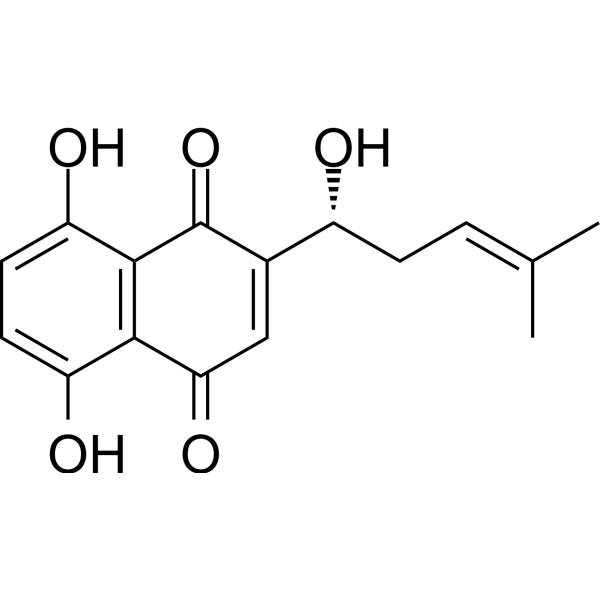
    Shikonin

    紫草素

    Inhibitor 99.80%
    Shikonin 是中草药紫草的主要成分。Shikonin 是一种有效的 TMEM16A 氯化物通道 (chloride channel) 抑制剂,IC50 为 6.5 μM。Shikonin 是一种特异的丙酮酸激酶 M2 (PKM2) 抑制剂,还可以抑制 TNF-αNF-κB途径。Shikonin 通过抑制糖酵解降低外泌体 (exosome) 的分泌。Shikonin 抑制 AIM2炎性体活化。
    Shikonin
  • HY-P9970
    Infliximab

    英夫利昔单抗

    Inhibitor
    Infliximab (Avakine) 是一种与 TNF-α 特异性结合的嵌合单克隆 IgG1 抗体。Infliximab 可阻止 TNF-α 与 TNF-α 受体 1 (TNFR1) 和 TNFR2 的相互作用。Infliximab 可用于自身免疫,慢性炎症性疾病和糖尿病神经病变的研究。
    Infliximab
  • HY-N0182
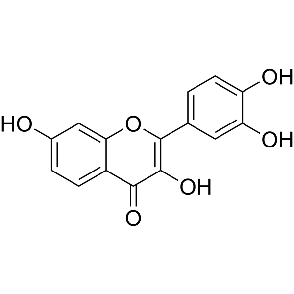
    Fisetin

    漆黄素

    Inhibitor 98.39%
    Fisetin是一种在许多水果和蔬菜中发现的天然黄酮醇,具有多种益处,如抗氧化,抗癌,神经保护作用。
    Fisetin
  • HY-P9908
    Adalimumab

    阿达木单抗

    Inhibitor 99.62%
    Adalimumab 是一种人源的单克隆 IgG1 抗体,靶向肿瘤坏死因子α (TNF-α)。
    Adalimumab
  • HY-P990007
    Tulisokibart Inhibitor
    Tulisokibart (PRA023) 是人源化后的小鼠 IgG1-κ 型抗体,靶向 TNFSF15/TL1A。Tulisokibart 可用于多种炎症/纤维化疾病的研究,例如克罗恩病 (Crohn's Disease, CD)。
    Tulisokibart
  • HY-P990008
    Atrosab Antagonist
    Atrosab 是一种人源化 IgG1 拮抗性抗 TNFR1 抗体。Atrosab 抑制 TNF 介导的细胞凋亡 (apoptosis) 以及 IL-6 和 IL-8 的产生。Atrosab 可用于炎症性疾病的研究。
    Atrosab
  • HY-P99796
    Ozoralizumab Inhibitor 99.80%
    Ozoralizumab (ATN-103) 是一种抗 TNFα 人源化抗体。Ozoralizumab 是一种人源化三价纳米抗体化合物,由两个抗人 TNFα 纳米抗体和一个抗人血清白蛋白 (HSA) 纳米抗体组成。Ozoralizumab 可用于关节炎的研究。
    Ozoralizumab
  • HY-108847
    Etanercept

    依那西普

    Inhibitor 99.20%
    Etanercept 是一种结合肿瘤坏死因子 (TNF) 的二聚体融合蛋白,作为 TNF 抑制剂。Etanercept 竞争性地抑制 TNF-α 和 TNF-β 与细胞表面 TNF 受体的结合,使肿瘤坏死因子在生物学上失去活性。Etanercept 对风湿性关节炎、幼年特发性关节炎和斑块性银屑病有效。
    Etanercept
  • HY-P0224
    N-Formyl-Met-Leu-Phe Inhibitor 99.83%
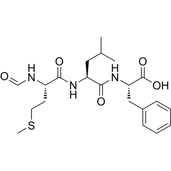
    N-Formyl-Met-Leu-Phe (fMLP; N-Formyl-MLF) 是一种趋化肽和N-甲酰基肽受体 (FPR) 的特异性配体。报道显示N-Formyl-Met-Leu-Ph 可抑制 TNF-alpha 的分泌。
    N-Formyl-Met-Leu-Phe
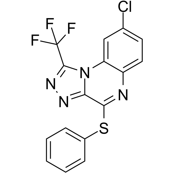
  • HY-13812
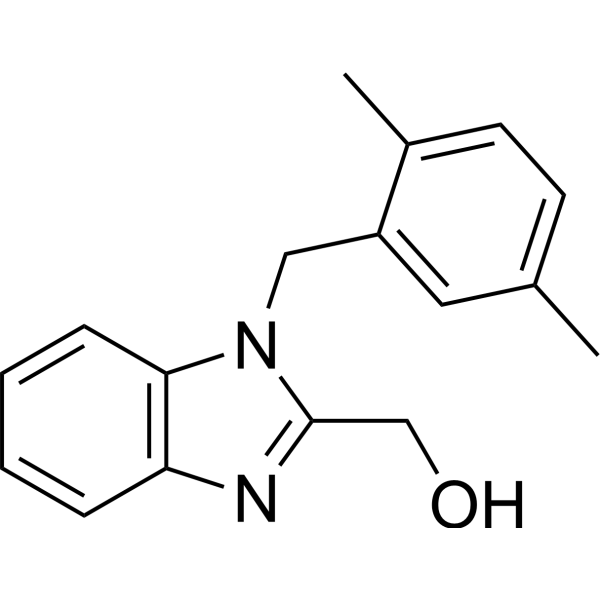
    QNZ Inhibitor 99.51%
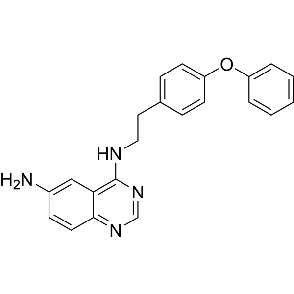
    QNZ (EVP4593) 强抑制 NF-κB 转录激活和 TNF-α 产生,IC50 分别为 11 和 7 nM。QNZ (EVP4593) 是一种保护神经的钙池操纵的钙通道 (SOC) 抑制剂。
    QNZ
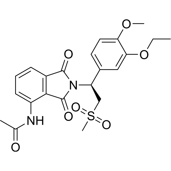
  • HY-12085
    Apremilast

    阿普司特

    Inhibitor 99.91%
    Apremilast (CC-10004) 是一种口服有效的磷酸二酯酶 4 (PDE4) 抑制剂,IC50 为 74 nM。Apremilast 抑制脂多糖 (LPS) 释放 TNF-α,IC50 为 104 nM。
    Apremilast
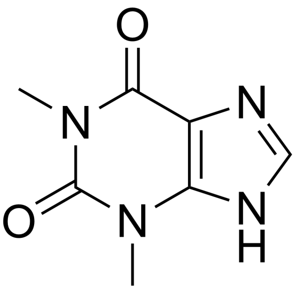
  • HY-B0809
    Theophylline

    茶碱

    Inhibitor 99.89%
    Theophylline (1,3-Dimethylxanthine) 是有效的磷酸二酯酶 (PDE) 抑制剂,腺苷受体拮抗剂,和组蛋白脱乙酰酶 (HDAC) 活化剂。Theophylline (1,3-Dimethylxanthine) 抑制 PDE3 活性,放松气道平滑肌。Theophylline (1,3-Dimethylxanthine) 通过增加 IL-10 和抑制 NF-κ B进入细胞核而具有抗炎活性。Theophylline (1,3-Dimethylxanthine) 诱发细胞凋亡 (apoptosis)。Theophylline (1,3-Dimethylxanthine) 可用于哮喘和慢性阻塞性肺疾病 (COPD) 的研究。
    Theophylline
  • HY-110203
    R-7050 Antagonist 98.55%
    R-7050 (TNF-α Antagonist III) 是一种肿瘤坏死因子受体 (TNFR) 拮抗剂,对 TNFα 具有更高选择性。
    R-7050
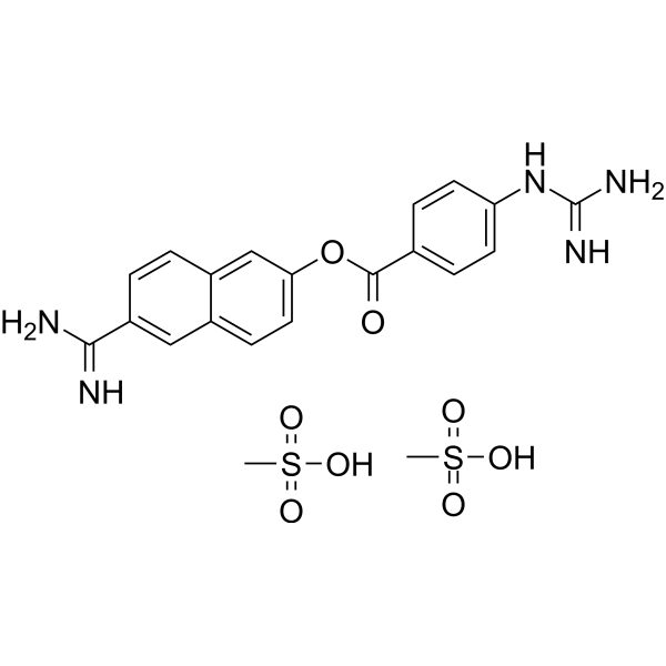
  • HY-B0190A
    Nafamostat mesylate

    甲磺酸萘莫司他

    98.06%
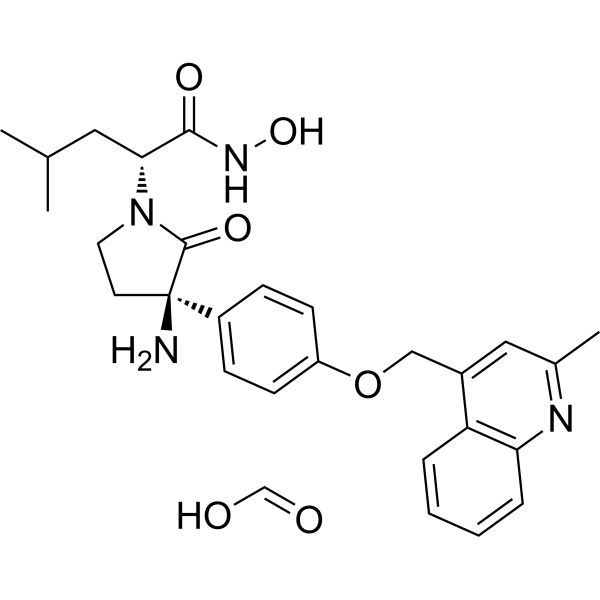
    Nafamostat mesylate (FUT-175),一种抗凝剂,是一种人工合成的丝氨酸蛋白酶 (serine protease) 抑制剂。 Nafamostat mesylate 具有抗癌和抗病毒作用,Nafamostat mesylate 通过上调肿瘤坏死因子受体 1 (TNFR1) 的表达诱导凋亡 (apoptosis),可用于动脉壁病理性增厚的发生发展3]
    Nafamostat mesylate
  • HY-120934
    C25-140 Inhibitor 99.84%
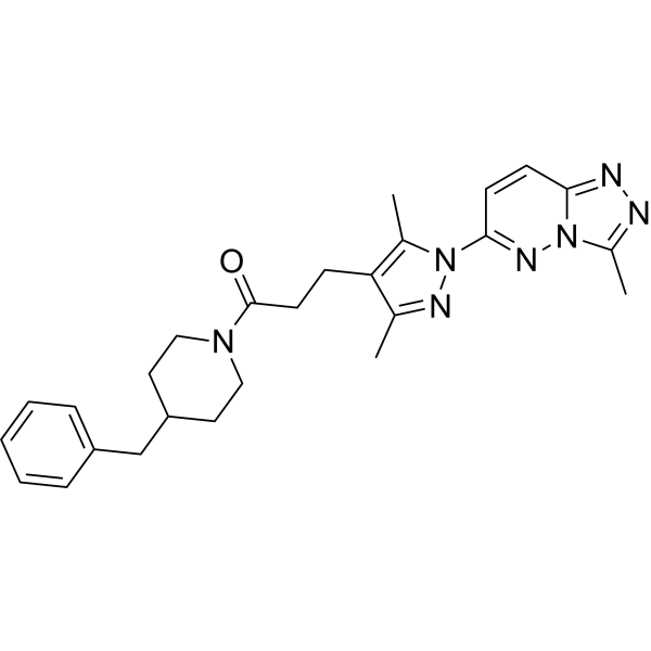
    C25-140 是一种首创的,具有口服活性和一定选择性的 TRAF6-Ubc13 相互作用的抑制剂,直接与 TRAF6 结合,阻断 TRAF6 和 Ubc13 的相互作用,从而降低 TRAF6 活性,降低 NF-κB 的活性,并对抗自身免疫。
    C25-140
  • HY-123942
    Diprovocim Agonist 98.35%
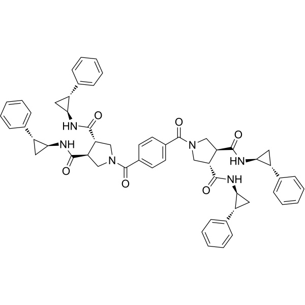
    Diprovocim 是一种有效的 TLR1/TLR2 激动剂。Diprovocim 在人 THP-1 细胞中引发完全激动剂活性 (EC50=110 pM)。Diprovocim 刺激小鼠巨噬细胞释放 TNF-α (EC50=1.3 nM)。Diprovocim 激活下游 MAPKNF-κB 信号通路。Diprovocim 在小鼠中显示出很强的佐剂活性,尤其是促进细胞免疫反应。
    Diprovocim
  • HY-133122
    UCB-9260 Inhibitor 99.96%
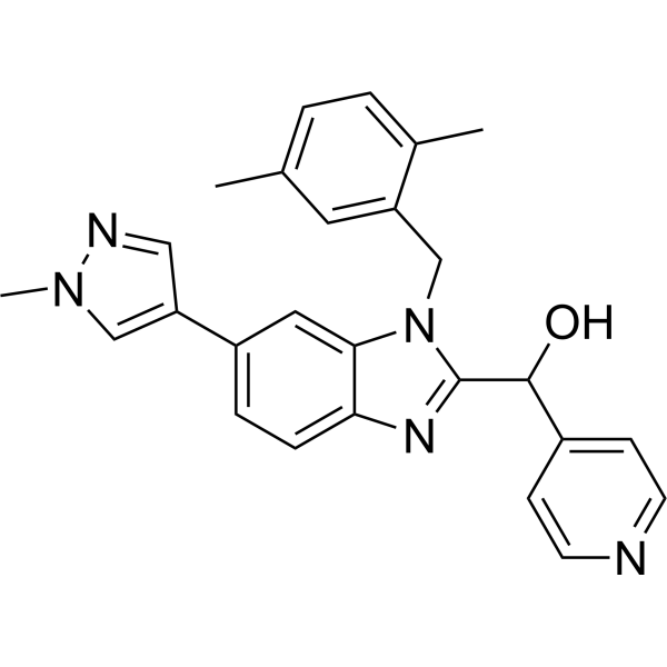
    UCB-9260 是一种口服活性小分子,通过稳定三聚体的不对称形式抑制肿瘤坏死因子 (TNF) 信号传导。UCB-9260 对肿瘤坏死因子的选择性高于其他超家族成员,并与肿瘤坏死因子结合,其 Kd 值为 13 nM。
    UCB-9260
  • HY-P99107
    Brentuximab vedotin

    维布妥昔单抗; 本妥昔单抗

    Inhibitor 98.00%
    Brentuximab vedotin (cAC10-vcMMAE) 是一种抗体-活性分子偶联物 (ADC),由抗 CD30 抗体和 Monomethyl auristatin E (MMAE) 组成。Brentuximab vedotin 抑制 CD30 阳性细胞,其 IC50 值为 2.5 ng/mL。Brentuximab vedotin 可用于复发难治性霍奇金淋巴瘤的研究。
    Brentuximab vedotin
  • HY-N0633
    Muscone

    麝香酮

    Inhibitor ≥98.0%
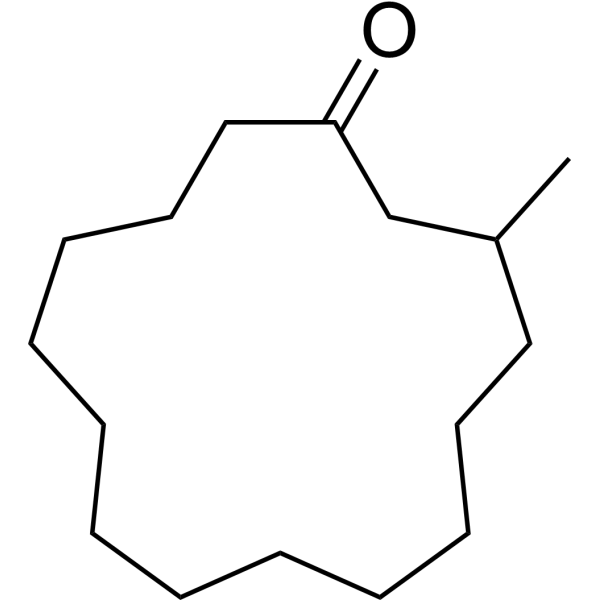
    Muscone 是中药麝香的主要活性单体。Muscone 抑制 NF-κBNLRP3 炎性小体的活化。Muscone 显着降低炎性细胞因子 (IL-1βTNF-αIL-6) 水平,并最终改善心脏功能和存活率。
    Muscone
目录号 产品名 / 同用名 种属 表达系统

Following the binding of TNF to TNF receptors, TNFR1 binds to TRADD, which recruits RIPK1, TRAF2/5 and cIAP1/2 to form TNFR1 signaling complex I; TNFR2 binds to TRAF1/2 directly to recruit cIAP1/2. Both cIAP1 and cIAP2 are E3 ubiquitin ligases that add K63 linked polyubiquitin chains to RIPK1 and other components of the signaling complex. The ubiquitin ligase activity of the cIAPs is needed to recruit the LUBAC, which adds M1 linked linear polyubiquitin chains to RIPK1. K63 polyubiquitylated RIPK1 recruits TAB2, TAB3 and TAK1, which activate signaling mediated by JNK and p38, as well as the IκB kinase complex. The IKK complex then activates NF-κB signaling, which leads to the transcription of anti-apoptotic factors-such as FLIP and Bcl-XL-that promote cell survival. 


The formation of TNFR1 complex IIa and complex IIb depends on non-ubiquitylated RIPK1. For the formation of complex IIa, ubiquitylated RIPK1 in complex I is deubiquitylated by CYLD. This deubiquitylated RIPK1 dissociates from the membrane-bound complex and moves into the cytosol, where it interacts with TRADD, FADD, Pro-caspase 8 and FLIPL to form complex IIa. By contrast, complex IIb is formed when the RIPK1 in complex I is not ubiquitylated owing to conditions that have resulted in the depletion of cIAPs, which normally ubiquitylate RIPK1. This non-ubiquitylated RIPK1 dissociates from complex I, moves into the cytosol, and assembles with FADD, Pro-caspase 8, FLIPL and RIPK3 (but not TRADD) to form complex IIb. For either complex IIa or complex IIb to prevent necroptosis, both RIPK1 and RIPK3 must be inactivated by the cleavage activity of the Pro-caspase 8-FLIPL heterodimer or fully activated caspase 8. The Pro-caspase 8 homodimer generates active Caspase 8, which is released from complex IIa and complex IIb. This active Caspase 8 then carries out cleavage reactions to activate downstream executioner caspases and thus induce classical apoptosis. 


Formation of the complex IIc (necrosome) is initiated either by RIPK1 deubiquitylation mediated by CYLD or by RIPK1 non-ubiquitylation due to depletion of cIAPs, similar to complex IIa and complex IIb formation. RIPK1 recruits numerous RIPK3 molecules. They come together to form amyloid microfilaments called necrosomes. Activated RIPK3 phosphorylates and recruits MLKL, eventually leading to the formation of a supramolecular protein complex at the plasma membrane and necroptosis [1][2].


Reference:
[1]. Brenner D, et al. Regulation of tumour necrosis factor signalling: live or let die.Nat Rev Immunol. 2015 Jun;15(6):362-74. 
[2]. Conrad M, et al. Regulated necrosis: disease relevance and therapeutic opportunities.Nat Rev Drug Discov. 2016 May;15(5):348-66.