1. Autophagy
  2. Autophagy
  3. Hoechst 33342

Hoechst 33342  (Synonyms: bisBenzimide H 33342; HOE 33342)

目录号: HY-15559 纯度: 99.94%
COA 产品使用指南

Hoechst 33342 是 Hoechst 系列的标记染料。Hoechst 系列是活细胞核标记染料。Hoechst 通过结合 DNA 双链中的小沟而结合核酸。Hoechst 倾向于结合富含 A/T 的 DNA 链。同时,富含 A/T 的双链 DNA 使荧光强度显著增强。Hoechst 可穿过细胞膜,结合活细胞或固定细胞。Hoechst 染料的荧光强度随着溶液 pH 升高而增强。

MCE 的所有产品仅用作科学研究或药证申报,我们不为任何个人用途提供产品和服务

Hoechst 33342 Chemical Structure

Hoechst 33342 Chemical Structure

CAS No. : 23491-52-3

1.  客户无需承担相应的运输费用。

2.  同一机构(单位)同一产品试用装仅限申领一次,同一机构(单位)一年内

     可免费申领三个不同产品的试用装。

3.  试用装只面向终端客户

规格 价格 是否有货 数量
25 mg ¥500
In-stock
50 mg ¥800
In-stock
100 mg ¥1200
In-stock
200 mg   询价  
500 mg   询价  

* Please select Quantity before adding items.

Customer Review

Other Forms of Hoechst 33342:

MCE 顾客使用本产品发表的 53 篇科研文献

IF

    Hoechst 33342 purchased from MCE. Usage Cited in: Bioact Mater. 2022 Mar 17;18:91-103.  [Abstract]

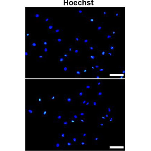
    The fluorescent images of HUVECs stained with NO fluorescent probe (DAF-FM DA) and Hoechst 33342 after being treated with UCATS under dark conditions or 808 nm NIR irradiation.

    Hoechst 33342 purchased from MCE. Usage Cited in: Chem Eng J. 15 December 2022, 138004.

    Apoptotic cells in four groups were stained with Hoechst 33342 (for 10 min) reflected by bright blue nuclei, including hP-MSCs cultured on noncoated, L-Gel-coated (10 nM), and D-Gel coated (10 nM) plates under exposure of 500 μM H2O2 for 5 h except for the normal group, and PBS as control.

    Hoechst 33342 purchased from MCE. Usage Cited in: J Lipid Res. 2022 May;63(5):100207.  [Abstract]

    complete medium containing 1 μg/mL BODIPY 500/510 for 3 h and then 2 μg/mL Hoechst 33342 for an additional 2 min.

    Hoechst 33342 purchased from MCE. Usage Cited in: Theranostics. 2021 Mar 4;11(10):4655-4671.  [Abstract]

    Hoechst 33342 (10 µg/mL) is added to the medium of cultured megakaryocytes for 2 h at 37°C. Cells are stained with the directly coupled monoclonal antibodies PE-anti-CD41 and APC-anti-Mpl for 30 min at 4°C. Ploidy is measured in the CD41+Mpl+ cell population.

    Hoechst 33342 purchased from MCE. Usage Cited in: J Cell Biol. 2021 Oct 4;220(10):e202011153.  [Abstract]

    Chromatin is briefly counterstained with Hoechst 33342 (5 µg/mL) before imaging.

    Hoechst 33342 purchased from MCE. Usage Cited in: Aging Cell. 2021 Jul;20(7):e13382.  [Abstract]

    BM cells are incubated with 5 μg/mL Hoechst 33342 in Hank balanced salt solution containing 20 mM HEPES, 5 mM glucose, and 10% fetal bovine serum at 37°C for 45 min, followed by an additional 45 min of incubation with 1 μg/ml Pyronin Y.

    Hoechst 33342 purchased from MCE. Usage Cited in: Stem Cell Res Ther. 2021 Jan 22;12(1):77.  [Abstract]

    To evaluate the anti-apoptosis property of extracellular vesicles from hP-MSCs, the Hoechst 33342 (for 10 min) is used to distinguish the apoptotic cells in the three different groups.

    Hoechst 33342 purchased from MCE. Usage Cited in: Acta Biomater. 2020 Sep 1;113:289-304.  [Abstract]

    Hoechst 33342 staining (for 6 h) is used to distinguish apoptotic changes in H2O2-treated hP-MSCs.

    Hoechst 33342 purchased from MCE. Usage Cited in: ACS Appl Mater Interfaces. 2018 Sep 12;10(36):30081-30091.  [Abstract]

    Hoechst 33342 staining shows the survival of HUVECs administrated 100 μg/mL exosomes or CS-Exo upon exposure to 400 μM H2O2.

    Hoechst 33342 purchased from MCE. Usage Cited in: Chinese Pharmacological Bulletin. 2018 May; 34(5): 620-626.

    PC12 cells are treated with 1.5 mM Methylglyoxal (MG) for 24 h before pretreatment with Butein (2.5, 5 μM) for 1 h. Propidium iodide (PI) and Hoechst 33342 are used to determine cell apoptosis.
    • 生物活性

    • 实验参考方法

    • 纯度 & 产品资料

    • 参考文献

    生物活性

    Hoechst 33342 is a marker dye in Hoechst series. Hoechst is A live nuclear marker dye. Hoechst binds to the grooves in the DNA double strand, which tends to be A/ T-rich DNA strand. Although it binds to all nucleic acids, the A/ T-rich double strand DNA significantly enhances fluorescence intensity Therefore,Hoechst dye can be used for living cell labeling. The fluorescence intensity of Hoechst dye increases with the increase of pH of solution[1].

    IC50 & Target

    Dye reagent[1]
    DNA Stain[1]

    体外研究
    (In Vitro)

    Hoechst 工作液的配制
    1.1 制备储存液
    用 DMSO 配制 1 mg/mL 的 Hoechst 储存液。
    注:Hoechst 储存液建议分装后于 -4℃ 或 -20℃ 避光保存。
    1.2 工作液的配制
    用预热好的无血清细胞培养基或 PBS 稀释储存液,终浓度为 10 μg/mL Hoechst 工作液。
    注:请根据实际情况调整 Hoechst 工作液浓度,且现用现配。
    2. 细胞染色(悬浮细胞)
    2.1 离心收集细胞,加入 PBS 洗涤两次,每次 5 分钟。细胞密度在 1×10⁶/mL
    2.2 加入 1 mL Hoechst 工作液,室温孵育 3-10 分钟。
    2.3 400 g,离心 3-4 分钟,弃去上清。
    2.4 加入 PBS 洗涤细胞两次,每次 5 分钟。
    2.5 用 1 mL 无血清培养基或 PBS 重悬细胞后,使用荧光显微镜或流式细胞仪进行观察。
    3. 细胞染色(贴壁细胞)
    3.1 将贴壁细胞培养于无菌盖玻片上。
    3.2 从培养基中移出盖玻片,吸除多余培养基。
    3.3 加入 100 μL 染料工作液,轻轻晃动使其完全覆盖细胞,孵育 3-10 分钟。
    3.4 吸去染料工作液,用培养基洗 2-3 次,每次 5 分钟,使用荧光显微镜或流式细胞仪进行观察。


    注意事项
    1. 请根据实际情况调整 Hoechst 工作液浓度。
    2. 本产品仅限于专业人员的科学研究用,不得用于临床诊断或治疗,不得用于食品或药品。
    3. 为了您的安全和健康,请穿实验服并戴一次性手套操作。

    MCE has not independently confirmed the accuracy of these methods. They are for reference only.

    分子量

    452.55

    Formula

    C27H28N6O

    CAS 号
    性状

    固体

    颜色

    Brown to breen

    Emission (Em)

    452

    Excitation (Ex)

    356

    运输条件

    Room temperature in continental US; may vary elsewhere.

    储存方式

    4°C, protect from light

    *In solvent : -80°C, 6 months; -20°C, 1 month (protect from light)

    溶解性数据
    细胞实验: 

    DMSO 中的溶解度 : 6.25 mg/mL (13.81 mM; 超声助溶 (<60°C); 吸湿的 DMSO 对产品的溶解度有显著影响,请使用新开封的 DMSO)

    H2O 中的溶解度 : < 0.1 mg/mL (insoluble)

    配制储备液
    浓度 溶剂体积 质量 1 mg 5 mg 10 mg
    1 mM 2.2097 mL 11.0485 mL 22.0970 mL
    5 mM 0.4419 mL 2.2097 mL 4.4194 mL
    查看完整储备液配制表

    * 请根据产品在不同溶剂中的溶解度选择合适的溶剂配制储备液;一旦配成溶液,请分装保存,避免反复冻融造成的产品失效
    储备液的保存方式和期限:-80°C, 6 months; -20°C, 1 month (protect from light)。-80°C储存时,请在6个月内使用,-20°C储存时,请在1个月内使用。

    • 摩尔计算器

    • 稀释计算器

    Mass (g) = Concentration (mol/L) × Volume (L) × Molecular Weight (g/mol)

    质量
    =
    浓度
    ×
    体积
    ×
    分子量 *

    Concentration (start) × Volume (start) = Concentration (final) × Volume (final)

    This equation is commonly abbreviated as: C1V1 = C2V2

    浓度 (start)

    C1

    ×
    体积 (start)

    V1

    =
    浓度 (final)

    C2

    ×
    体积 (final)

    V2

    动物实验:

    请根据您的 实验动物和给药方式 选择适当的溶解方案。

    以下溶解方案都请先按照 In Vitro 方式配制澄清的储备液,再依次添加助溶剂:
    ——为保证实验结果的可靠性,澄清的储备液可以根据储存条件,适当保存;体内实验的工作液,建议您现用现配,当天使用
    以下溶剂前显示的百分比是指该溶剂在您配制终溶液中的体积占比;如在配制过程中出现沉淀、析出现象,可以通过加热和/或超声的方式助溶

    • 方案 一

      请依序添加每种溶剂: 10% DMSO    40% PEG300    5% Tween-80    45% Saline

      Solubility: ≥ 0.5 mg/mL (1.10 mM); 澄清溶液

      此方案可获得 ≥ 0.5 mg/mL(饱和度未知)的澄清溶液。

      1 mL 工作液为例,取 100 μL 5.0 mg/mL 的澄清 DMSO 储备液加到 400 μL PEG300 中,混合均匀;再向上述体系中加入 50 μL Tween-80,混合均匀;然后再继续加入 450 μL 生理盐水 定容至 1 mL

      生理盐水的配制:将 0.9 g 氯化钠,溶解于 ddH₂O 并定容至 100 mL,可以得到澄清透明的生理盐水溶液。
    • 方案 二

      请依序添加每种溶剂: 10% DMSO    90% (20% SBE-β-CD in Saline)

      Solubility: ≥ 0.5 mg/mL (1.10 mM); 澄清溶液

      此方案可获得 ≥ 0.5 mg/mL(饱和度未知)的澄清溶液。

      1 mL 工作液为例,取 100 μL 5.0 mg/mL 的澄清 DMSO 储备液加到 900 μL 20% 的 SBE-β-CD 生理盐水水溶液 中,混合均匀。

      20% SBE-β-CD in Saline 的配制(4°C,储存一周):2 g SBE-β-CD(磺丁基醚 β-环糊精)粉末定容于 10 mL 的生理盐水中,完全溶解至澄清透明。
    动物溶解方案计算器
    请输入动物实验的基本信息:

    给药剂量

    mg/kg

    动物的平均体重

    g

    每只动物的给药体积

    μL

    动物数量

    由于实验过程有损耗,建议您多配一只动物的量
    请输入您的动物体内配方组成:
    %
    DMSO +
    +
    %
    Tween-80 +
    %
    Saline
    如果您的动物是免疫缺陷鼠或者体弱鼠,建议 DMSO 中的在最后工作液体系中的占比尽量不超过 2%。
    方案所需 助溶剂 包括:DMSO ,均可在 MCE 网站选购。 Tween 80,均可在 MCE 网站选购。
    计算结果
    工作液所需浓度 : mg/mL
    储备液配制方法 : mg 药物溶于 μL  DMSO(母液浓度为 mg/mL)。

    *In solvent : -80°C, 6 months; -20°C, 1 month (protect from light)

    您所需的储备液浓度超过该产品的实测溶解度,以下方案仅供参考,如有需要,请与 MCE 中国技术支持联系。
    动物实验体内工作液的配制方法 : 取 μL DMSO 储备液,加入 μL  μL ,混合均匀至澄清,再加 μL Tween 80,混合均匀至澄清,再加 μL 生理盐水
    连续给药周期超过半月以上,请谨慎选择该方案。
    请确保第一步储备液溶解至澄清状态,从左到右依次添加助溶剂。您可采用超声加热 (超声清洗仪,建议频次 20-40 kHz),涡旋吹打等方式辅助溶解。
    纯度 & 产品资料

    纯度: 99.94%

    染色示例
    参考文献
    Cell Assay
    [1]

    Labeling Nuclear DNA with Hoechst 33342[1] Step 1, Dilute the Hoechst stock solution 1:100 in H2O for use in labeling. Step 2, Aspirate the cell medium from cells grown on coverslips. Rinse the cells three times with PBS+. Step 3, Incubate the cells in the Hoechst labeling solution (from Step 1) for 10-30 min at room temperature. Step 4, Aspirate the labeling solution. Rinse the cells three times in PBS+. Step 5, Mount the coverslips. Step 6, Image the cells (λex ~353 nm, λem ~483 nm for Hoechst 33342)[1].

    MCE has not independently confirmed the accuracy of these methods. They are for reference only.

    参考文献

    完整储备液配制表

    * 请根据产品在不同溶剂中的溶解度选择合适的溶剂配制储备液;一旦配成溶液,请分装保存,避免反复冻融造成的产品失效
    储备液的保存方式和期限:-80°C, 6 months; -20°C, 1 month (protect from light)。-80°C储存时,请在6个月内使用,-20°C储存时,请在1个月内使用。

    可选溶剂 浓度 溶剂体积 质量 1 mg 5 mg 10 mg 25 mg
    DMSO 1 mM 2.2097 mL 11.0485 mL 22.0970 mL 55.2425 mL
    5 mM 0.4419 mL 2.2097 mL 4.4194 mL 11.0485 mL
    10 mM 0.2210 mL 1.1049 mL 2.2097 mL 5.5243 mL
    Help & FAQs
    • Do most proteins show cross-species activity?

      Species cross-reactivity must be investigated individually for each product. Many human cytokines will produce a nice response in mouse cell lines, and many mouse proteins will show activity on human cells. Other proteins may have a lower specific activity when used in the opposite species.

    您最近查看的产品:

    Your information is safe with us. * Required Fields.

       产品名称:


    * 需求量:

    * 客户姓名:


    * Email:

    * 电话:


    * 公司或机构名称:

       留言给我们:

    Bulk Inquiry

    Inquiry Information

    产品名称:
    Hoechst 33342
    目录号:
    HY-15559
    需求量: